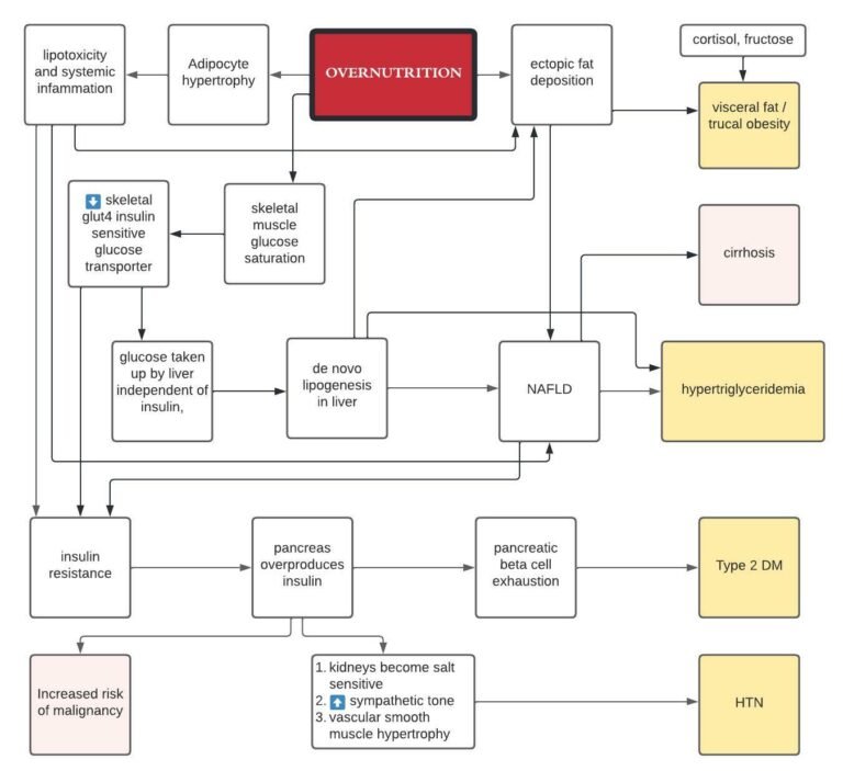
Why do we have to die?

If we are to understand aging and death, the primary question is why should there be aging and death in the first place?  What evolutionary purpose might they serve?  First let me say that I’m discouraged that I don’t have any formal education in evolutionary biology, I think it’s a fascinating field and I’m frustrated by my status as a dilettante.  Yet I proceed in the spirit of an amateur.

Established evolutionary theory holds that genes that lead to the death of an organism would not be adaptive and so aging and death are not part of some program.  Current accepted theories fall under two broad categories, both animated by the spirit that aging is a product of evolutionary neglect, not intent.

The first theory is that of “accumulated mutations,” which holds that the organism accrues wear and tear from the processes of life, primarily genetic mutations that compromise function and lead to aging and death.  Given the evolving understanding of genetics as well as our new awareness that the hallmarks of aging extend beyond simple genetic mutations, this is theory is now a non-starter.

The second theory that is typically offered is “antagonistic pleiotropy,” meaning that survival to reproduction is prioritized by evolution and the very genes that improve survival to reproductive age lead to aging and death with the passage of time.  Or put differently, genes that code to preserve the organism do not improve fitness.  This theory is of a different class than the first one—while the first one is mechanistic and has been disproven, this one is teleological, offering an unverifiable story.

Neither of these theories supports the idea that aging and death are evolved and part of some program.  I’m suggesting that the field of evolutionary biology is wrong and I want to argue instead that dying is a way to promote evolutionary fitness.  Let me just make it clear:  I believe that aging is an extension of the same program as development and this program has evolved because it increased iterations of natural selection and evolutionary fitness.

The only way I can make this point is with a story, so let us consider the example of a nonhuman organism, an early mammal, though we could just as well pick a reptile or something else that is subject to sexual reproduction.  Also one presumption–that a given population lives in a particular geography with limited natural resources.

Consider first the non-aging organism.  At a certain point, the mature male will be competing for mating partners with males of the next generation.  Given the evolutionary imperative to pass on genes, there will be competition, and since our mature male is larger and more experienced than the next generation, it will kill them.  Alternatively, it will simply scare them off and prohibit them from mating.  But killing them is better as it will prevent them from getting larger one day and becoming a legitimate adversary.  Which is to say, those populations that evolve to infanticide will be better off.  So now you have an immortal infanticidal creature that continues to mate forever.  And if there is an accident or some lethal event, perhaps the accumulation of damage, the younger offspring can finally take his place.  But you have a real limit on the amount of natural selection because the population genetics are all deriving from the same male, who is presumably the one mating with all the females.

On the other hand, in an aging organism, the old male always gets weaker with time and is superseded by the young males, who fight it out for the right to mate with the females.  Because there is death, this arrangement maximizes iterations of natural selection which selects for better evolutionary fitness.  Invariably this population will evolve more rapidly and will likely be better suited to survive in a difficult environment.

So aging and death can be considered an evolutionary advantage.  Why couldn’t it be this simple?

Circadian interventions

We have internal rhythms that align with the rotation of the planet.  Most famously, there is the rhythmic secretion of melatonin at night and a spike of cortisol in the morning.  These and myriad other oscillations are controlled by aptly named clock genes that are present in every cell of the body. Clock genes work together to maintain and synchronize the timing of various physiological and behavioral processes in the human body. Some of the key clock genes are CLOCK, BMAL1, PER, and CRY.   The molecular clock in individual cells is synchronized by a master oscillator located in the suprachiasmatic nucleus (SCN) of the hypothalamus. The SCN receives light signals from the retina, which helps to align the internal clock with the external light-dark cycle, ensuring that the circadian rhythm is properly entrained to the external environment.  Circadian dysfunction that occurs when peripheral clocks are out of sync with the central clock, as during jet lag or shift work, can lead to metabolic dysfunction. The function of the clock genes and the mechanisms of circadian rhythmicity have been elucidated to fascinating detail–though their translation to clinical medicine has not happened.  But according to this Montenegrin paper, circadian timing can be easily applied for interventions related to metabolic health.

Certainly the most obvious and best studied of the circadian recommendations is to align sleep with the day and night cycles.  Following that, eating while the sun is out is a good rule of thumb, we know that eating outside a circadian window affects how we use our calories and increases the likelihood of metabolic dysfunction.  Like many who practice obesity medicine, I suggest front loading calories earlier in the day.

Exercise in the morning–I’ve read different perspectives, but the paper makes a good case for timing exercise early.  They point out that melatonin phase delays are associated with exercise in the evening or overnight.

Antihypertensives:  ACE inhibitors and ARB’s should be taken in the evening–I didn’t know this.

Statins:  Should also be taken at night.  Good to know.  Put it on your nightstand.

Metformin:  take it at night

Breakfast:  Is it the most important meal of the day or should you skip breakfast to prolong your fast?  It appears that calories eaten for breakfast are less fattening, account for more energy expenditure and may reinforce important clock genes.  Which is to say, eat your breakfast.

GLP-1 RA’s in the afternoon to sync with endogenous GLP-1 secretion–probably less relevant since people take once weekly dosing.

Why Nature Wants Us to be Fat by Richard Johnson

The two popular theories that purport to explain the obesity pandemic are the carbohydrate insulin model and the standard model of energy balance.  In Why Nature Wants Us to be Fat, nephrologist Richard Johnson bravely proposes another theory:  the survival switch.

In his formulation, nature has evolved a survival mechanism allowing organisms to rapidly gain weight in response to stress or in preparation for periods of scarcity.  It is meant to be turned on occasionally in response to adversity, but our food environment has interacted with our evolutionary biology to turn the switch on continuously, leading to widespread obesity.  The survival switch is mediated by the molecule fructose, and Johnson pins much of the blame on pervasive consumption of high fructose containing foods and beverages which were introduced in the 1970’s.  Fructose metabolism triggers an intracellular starvation type response in the liver that ultimately leads to leptin resistance and diminished satiety, which then causes persistently disordered appetite.  Fructose causes obesity not because of its caloric or nutritive value, but rather by hijacking appetite.  Sucrose too is implicated, as it is broken down to equal parts fructose and glucose.

The book impressively draws from biochemistry and clinical science as well as comparative biology, evolutionary science, bio-anthropology and paleontology to synthesize a coherent theory to explain our current global metabolic perdicament.  According to Johnson, a classic example of the survival switch is mammalian hibernation.  To take that example, in preparation for winter the bear markedly ramps up its consumption of fructose-containing blueberries in order to build fat stores, which become a source of both calories and water during its hibernation.  Similarly, birds often switch from insect diets to fruit and nectar-filled diets as winter approaches in order to gain fat.  In this way animal and fruit-bearing plant life are in seasonal harmony.

The fundamental issue is that fructose metabolism simulates an intracellular energy deficit in a dose dependent fashion by rapidly diminishing intracellular ATP by 20-60%.  As a result, appetite and fat deposition are stimulated.  Fructose is known to cause central leptin resistance by hypothalamic inflammation, which would naturally increase appetite.  Additionally, in the presence of fructose, saturated fats are particularly obesogenic.  Finally, mitochondria suffer oxidative stress because of fructose.

In the absence of fructose, other stimuli such as high salt, dehydration and umami (glutamate) can also trigger the survival switch by pushing conversion of glucose to sorbitol and then to fructose through the polyol pathway. High glycemic carbs will  increase fructose production as well, and this is probably the number one way in which people are exposed to fructose today outside of supplemented HFCS.

According to Johnson, animals are drawn to salt-licks because the salt triggers the conversion of glucose to fructose, which in turn helps them put on weight. To the extent that ultra-processed foods are formulated with high concentrations of salt and sugar, they are continually triggering this metabolic switch on a global scale.

Johnson suggests inhibitors of fructose metabolism would prevent the adverse consequences of the western diet and would by themselves prevent NAFLD.  There happens to be a number of Pharma groups working on fructokinase inhibitors.  Luteolin is a naturally occurring flavone that blocks fructose metabolism and has been associated with renal protection.

This is a fascinating book by a researcher who has stature and standing in the field, but his identification of the survival switch as the cause of the majority of  maladies afflicting mankind gives pause.    Yet the theory is cohesive, supported by empirical evidence and powerfully explains much of our current predicament.  I am swayed.

Muscle, Protein and Aging

Muscle is a metabolically active tissue that plays a central role in health and longevity.  Beyond functional roles such as helping one get up from a chair, going up stairs and avoiding falls as we age, muscle accounts for 75% of glucose disposal and makes the greatest contribution to resting energy expenditure of any tissue in the body.  For these reasons, we need to be mindful of preserving our muscle mass if we are to age well.  Unfortunately, it becomes harder to maintain (let alone build) muscle as we get older.  There is evidence of anabolic resistance in the elderly, a barrier to building muscle, and it is not uncommon for the elderly become sarcopenic.  Sarcopenia (from the Greek “loss of flesh”) can be a tremendous burden on geriatric populations and is the leading cause of frailty and deterioration.  What follows is a discussion of muscle metabolism that owes a debt of gratitude to the food industry scientist Don Layman and also to a review article by Tezze et. al.

Protein
The main drivers of muscle synthesis and retention are resistance exercise and to a lesser extent, protein intake.   But what kind of exercise?  And what kind of protein?  And how much?  And when?

Not all protein is equal.  

  • Protein is made of amino acids, some of which are categorized as essential because they cannot be manufactured by our body and must be consumed.
  • The essential amino acids are: histidine, isoleucine, leucine, lysine, methionine, phenylalanine, threonine, tryptophan and valine.
    • Leucine in particular is thought to be a notable driver of muscle protein synthesis
    • Leucine is also a primary driver of the enzyme mtorc1.
  • Proteins differ in their amino acid composition and degree of binding / accessibility.
  • Typically, meats and fish have a complete assortment of essential amino acids that are easily accessible for use in the body.
  • All essential amino acids are derived from bacteria.
    • Plants will absorb them as part of their root system to incorporate into plant protein.
    • Ruminative animals like cows have nitrogen fixing bacteria in their stomachs that very efficiently convert plant protein in grass / grain to essential amino acids, accounting for the high protein of herbivores who themselves consume very little protein.
  • Plants are often deficient in various essential amino acids and plant protein is more likely to present difficulties with extraction. They have a lower protein digestibility corrected amino acid score.
      • DIAAS is the digestible indispensable amino acid score.
      • To take an example, whey protein, a byproduct of cheesemaking, is 20% better than a soy protein isolate on the basis of its essential amino acids

Muscle Homeostasis

  • Muscle tissue is in a constant state of turnover characterized by rates of muscle protein breakdown and muscle protein synthesis.
  • The relationship of building muscle tissue to fostering longevity is seemingly paradoxical.  On the one hand, having muscle mass is good for aging and associated with less frailty.  On the other hand, it requires the anabolic activation of mtorc1, which is a driver of aging and frailty.  How do we reconcile these realities?
    • Muscle protein breakdown is inhibited by insulin, which is the body’s main anabolic hormone.
    • Beyond that, muscle protein synthesis is more heavily regulated and amenable to modification, typically stimulated by agents that increase the activity of mtor, which is believed to accelerate aging. We know that branched chain amino acids such as leucine drive mTORC1 and drive muscle growth.
    • Yet ITP studies in mice suggest L-leucine enriched diets reduce lifespan (ITP 2017) and chronic activation of mTORC1 stimulates progressive muscle damage and loss. Moreover, inhibition of mTORC1 with rapamycin prevents age-related muscle loss (1).
  • The resolution of this paradox is lies in the complexity of muscle homeostasis and the mTORC1 enzyme and there are various explanations.  Some authors (1) have suggested that Akt dependent activation of mTORC1 prevents muscle atrophy whereas Akt independent pathways will induce muscle atrophy, largely mediated by mitochondrial oxidative stress.  Others (2) have suggested that mTORC1 inhibition rejuvenates muscle stem cells by preventing senescence.

Another way of looking at this issue is to consider the essential amino acid leucine, which is a relatively muscle-specific activator of mtor.  Insulin on the other hand activates mtor in a range of tissues.  What this means functionally is that people who eat a lot of small carbohydrate meals, each of which leads to the secretion of insulin, continuously activate mTOR and drive anabolic pathways / aging across tissues.  This is probably the worst-case scenario and a real justification not to snack.   On the other hand, infrequent / periodic high protein meals with large leucine loads will stimulate muscle growth while reducing broader mtor activation associated with snacking.  Does this mean there is no role for carbs in the diet?  Of course, not, but that is a matter for another time.

Timing of protein consumption—is it better to spread your intake across the day or consume the bulk of your protein in one or two meal?

  • To build muscle you need large individual servings of protein.  Muscle anabolism is not stimulated until you have a significant ingestion. Muscle tissue senses the concentration of amino acids in the blood that serve as a signal that a meal has adequate quality for muscle to trigger the very expensive process of protein synthesis.
  • Protein should be front loaded in the day, ~30g for breakfast to interrupt overnight catabolism and then at dinner to forestall overnight catabolism. Ideally, we pair this with some resistance activity.
  • For the rest of your physiological needs (cardiac, liver, etc.), you don’t need large individual servings.
  • This is not true for kids—their muscle growth is more insulin oriented. They can use protein regardless of the absorption schedule.

Exercise and muscle

  • Resistance exercise is a necessary co-factor for muscle growth along with protein.
    • Resistance exercise stresses the muscle, disrupting homeostasis and leading to protein turnover. With protein turnover, there is protein misfolding, leading to endoplasmic reticulum stress.
    • ER stress leads to the activation of the “integrated stress response” a signaling pathway whose goal is to restore muscle homeostasis.
    • How? via upregulation of ATF4, which controls LARS, a leucine sensor.  Clearly more complicated than this, but here’s your broad outline.
  • Exercise induces both hyperplasia and hypertrophy of muscle fibers. In other words, it increases both the number of fibers and their size.
  • Exercise enhances the ability of the muscle to take-up more amino acids from the bloodstream, and this effect lasts multiple days.

Protein as a diet food

  • Protein is also the most satiating food.
  • It also has a higher thermogenic effect (burns more calories) than either carbs or fat. So eating protein it increases energy consumption.
  • Hi protein percentages during dieting preserves muscle mass.

Protein use / reversing catabolism with age

  • As we get older, our ability to use protein is diminished, referred to as “anabolic inflexibility.”
  • This is partly due to diminished physical activity with age, but also because of features intrinsic to the muscle tissue.
  • This anabolic resistance can be largely overcome by increasing protein intake.  Yet you also need resistance–it doesn’t matter how much protein you consume if you don’t also engage in some degree of muscle exercise. You need both.
  • Older adults need more protein.  There are varying recommendations between, 1-1.5 mg/kg/day (3), keeping in mind that not all protein is created equal.
  • I would rather that an older adult work hard to ensure adequate protein intake, eat meat fish and eggs every day, even if it means taking a statin for dyslipidemia. Obviously, this would be a case-by-case decision, but I’m far less interested in treating obesity, for instance in an older adult.

Cautionary thought:

  • Beyond driving muscle synthesis, certain branched chain amino acids found in animal sources such as isoleucine and isoleucine may actually drive mtor and accelerate aging.  In which case, in the setting of tonically activated mtor (middle/old age), a diet high in protein from vegetable sources makes most sense.
  1. https://www.ncbi.nlm.nih.gov/pmc/articles/PMC6738401/?fbclid=IwAR3vAqW1MLIF-kY1utROauhs2i8xHFSlqjeL95SRN02Cdgoif-FW1PWXw0c
  2. https://www.mikhailblagosklonny.com/blog/how-rapamycin-prevents-muscle-loss-and-sarcopenia-first-draft/
  3. https://pubmed.ncbi.nlm.nih.gov/24039411/

Metabolic Syndrome

The diagram below details how overfeeding will lead to the so-called metabolic syndrome, a cluster of conditions that are linked and can lead to vascular disease, stroke and heart attacks.  The components of  metabolic syndrome are hypertriglyceridemia, truncal obesity, hypertension, diabetes and low HDL.  This diagram elaborates an insulin-centric model, and it’s important to recognize that there are other models.

The overfed state means the cells of the body–primarily muscle, which accounts for 80% of the body’s glucose disposal, and adipose tissue–have been overwhelmed with fuel and become overstuffed.  Insulin is an important player in this schema because glucose transport into muscles requires insulin to get nutrition into the cell.  When the cells are full, they reduce their expression of insulin-dependent glut4 transporters, making it harder to push glucose into the cell.  This resistance can be overcome by manufacturing more insulin, which is how to body initially responds to the problem of rising glucose concentrations in the extra-cellular space.  Imagine insulin to be the hose pressure pushing gas into the tank.  The more stuffed the cells are, the more insulin you need to drive nutrition intracellularly, and this is precisely the phenotype of insulin resistance.  The pancreas is the hose pump in this analogy, and with prolonged pressure, it can fail leading to diabetes.  

Meanwhile glucose that should be going into the muscles and fat cells overflows from that compartment and is instead taken up by the liver in an insulin-independent process.  In the liver, glucose is converted to saturated fat through de novo lipogenesis, ultimately leading to non-alcoholic fatty liver disease (NAFLD), which can progress to cirrhosis and ultimately liver failure.  NAFLD has become the leading reason for referral to liver transplant centers.  

Meanwhile, the very high circulating levels of insulin cause sodium retention in the kidney, creating salt-sensitive hypertension.  At the same time, insulin acts more broadly as a growth factor in the body, and high levels of circulating insulin are associated with accelerated cell division and carcinogenesis.  

This schema is the backstory of the American healthcare system.  An ever rising percentage of patients in any emergency department have hypertension, hyperlipidemia, truncal obesity and diabetes.

Metabolic Flexibility

Humans evolved in the setting of periodic food scarcity during which we developed the ability to rapidly switch between different fuel sources.  We are designed by evolution to burn fat in our fasting state and to burn primarily carbohydrates in our fed state in order to generate energy in the form of ATP.  All of this occurs in our mitochondria.  But with the development of the modern industrial food environment and ready access to overnutrition combined with less energy expenditure compared to the conditions under which we evolved, we find ourselves overnourished and at odds with our evolutionary design.  One consequence of this mismatch is impaired fuel switching in the mitochondria—we lose the ability to easily switch between lipids and glucose that marks a healthy metabolism.  As a result, our engines are inefficient, we have trouble burning fat while fasting, we are hungrier sooner after a meal, we eat more and find ourselves in a feed forward loop.  Eventually we develop obesity and the diseases related to insulin resistance such as diabetes, metabolic syndrome, NAFLD and ultimately cancer.   At least these are the claims.  What follows is an abstract of a good review paper published earlier this week that examines the concept of metabolic flexibility.

How and why does metabolic inflexibility occur?  

With chronic overnutrition (aka the Standard American Diet), cells become overstuffed and congested with macronutrients, overwhelming the enzymatic mechanisms of metabolism and leading to accumulation of incompletely oxidized substrates–the details of this process are probably not as interesting for our purposes as are the consequences.  These substrates harm mitochondria through oxidative stress from free radical damage and acetylation / protein modification due to acetyl coA accumulation.  The damage actually reduces the number of mitochondria as well as changing their functional morphology–the mitochondria look different and their performance is degraded.   The plasticity of the fuel switch deteriorates, leading to a sluggish metabolic response to nutritional cues.  Imagine a hybrid vehicle whose gas motor is flooded and so it can only use electricity.  And maybe the gas tank is expansile and is ever filling.  (I wish I could think of a better metaphor but I’m a poor mechanic).

At the same time, the overfed state also requires increasing amounts of insulin to push glucose into cells via insulin dependent glut4 transporters.  Imagine insulin to be the hose pressure pushing gas into the tank.  The more stuffed the cells are, the more insulin you need to drive nutrition intracellularly, and this is precisely the phenotype of insulin resistance.  The pancreas is the hose pump in this analogy, and with prolonged pressure, it can fail leading to diabetes.  Meanwhile glucose that should be going into the muscles and fat cells is instead taken up by the liver in an insulin independent process and converted to fat through de novo lipogenesis, ultimately leading to NAFLD–now the leading cause of liver failure.  The very high circulating levels of insulin cause sodium retention in the kidney, creating the phenotype of salt-sensitive hypertension.  Insulin is also a growth factor, and high levels are associated with carcinogenesis.  This is the backstory of the American healthcare system.

Insulin resistance and metabolic inflexibility are intertwined and it’s not clear which comes first, but they relate to each other and are both a consequence of overfeeding.  One reason this topic is of clinical interest is that people with metabolic inflexibility have problems burning fat in the fasting state.  They get hungry sooner and have trouble with fasting.   The intracellular availability of glucose determines the nature of substrate oxidation in human subjects (1).  If there is an excess of glucose, the cell will preferentially oxidize the glucose.  Otherwise it will turn to fat.

How do we treat this metabolic rigidity?

  1. Diminish the nutrition going into the cell
    1. Caloric restriction: since the problem begins with too much energy, an energy deficit, regardless of macronutrient balance, is a reasonable first step toward relieving substrate competition.  I don’t think you need to go low carb or keto, though some people may find it easier to do so in order to manage their hunger.  Calorie restriction permits cells to decongest themselves of unused substrate and restore normal membrane potential across mitochondria.  Losing weight is the best and perhaps only valid treatment for fatty liver and can reverse insulin resistance and DM2 in its early stages.  So eating less is how to repair the metabolic inflexibility which derives from eating too much–it feels like an astute observation of the obvious, but there may be a special way of eating less that is especially effective.  Fasting.
    2. Fasting: starvation has benefits beyond calorie restriction alone.  We evolved to cope with periods of starvation and the metabolic program triggered by fasting has diverse benefits beginning with exhaustion of liver glycogen stores and initiation of ketosis. Burning ketones is metabolically healthy for a number of reasons, particularly as we age.  At the cellular level, fasting induces autophagy to remove damaged proteins, mitochondrial biogenesis and mitophagy.  The issue of fasting is, of course, a larger one and care must be taken to preserve lean muscle mass, but the rationale to starting a diet with a fast is that lipid oxidation is kickstarted by more rapidly inducing metabolic flexibility.
    3. SGLT2 inhibitors:  SGLT2 (sodium glucose cotransporter-2) inhibitors force the kidneys to spill glucose in the urine leading to a condition that mimics caloric restriction.  These drugs have been shown to be kidney protective, to reduce weight, to reduce blood pressure and to improve outcomes in congestive heart failure.  It turns out that they also promote beiging of adipocytes (in mice)—the brown fat that generates heat and burns calories in the process.  Unclear if this is relevant in humans.  At the mitochondrial level, SGLT2i’s permit the cell to decongest itself of the metabolic substrates that have caused all the damage.  Taking SGLT2i at night, according to the authors, amplifies the effects of an overnight fast.
  1. Improve factors that mitigate mitochondrial stress
    1. Restoration of glutathione with glyNAC to counter oxidant stress
    2. Carnitine based acetyl-group buffering: theoretical. I don’t see any evidence that carnitine supplementation is of value at this point.
  1. Increase energy expenditure
    1. Aerobic exercise, particularly zone 2 exercise is the best way to improve the number of mitochondria and their function. There is a strong association described between exercise and metabolic flexibility.  Even after one episode of exercise, insulin resistance and mitochondrial function improve.
    2. Resistance training: bigger muscles means a bigger glucose sink, a greater buffer to accommodate nutrient load in the fed state.


Footnote

(1) https://journals.physiology.org/doi/abs/10.1152/ajpendo.1996.270.4.e733

Autophagy

Autophagy (literally “self-eating”) is the body’s way of cleansing cells by recycling old or damaged components and is a process that appears to have a strong relationship to preventing disease and aging.  More technically, it is a highly regulated lysosome-dependent catabolic program used by the body to clear dysfunctional proteins, organelles and other structures that are typically first marked for the process by a protein called “ubiquitin.”  Primarily intra-cellular but also extra-cellular components are engulfed in autophagocytic vacuoles and degraded to constituent molecules, such as sugars, fatty acids and amino acids, which can be then be used as nutrition to produce ATP and/or provide building blocks for the synthesis of essential proteins.   There are different types of autophagy but for the most part they all serve this same purpose.  Mitophagy is a similar process that removes damaged or dysfunctional mitochondria.  There is also nucleophagy of material in the cell nucleus.


A number of types of autophagy are currently described.

  • Microautophagy: direct engulfment of cytoplasmic materials by invagination of lysosomal membrane.
  • Macroautophagy:  classic autophagasome mediated double membrane engulfment.
  • Chaperone Mediated autophagy: extremely selective target protein mediated (Hsc70) recycling that is activated in response to cellular stress.
  • Mitophagy:  autophagy related to mitochondria and their components
  • Also… ribophagy, lipophagy, xenophyagy, nucleophagy, clockophagy, lysophagy, etc.


Why is it important?
Autophagy is critical to cell function, regeneration and keeping the cell working properly.  Inhibition of autophagy by knocking out any of a variety of autophagy related genes invariably accelerates aging and disease.  Similarly, with aging, autophagy is downregulated (particularly in lymphocytes) leading to a forward feeding loop of accumulation of cellular debris and cellular dysfunction / dysregulation.   In contrast, enhanced autophagy delays aging and prolongs lifespan in a range of model organisms including yeast, nematodes, fruit flies and mice.  Lifespan studies in humans have not been conducted given the obvious challenges in conducting experiments in long-lived organisms, but it is generally assumed that regular induction of autophagy is a salubrious process, something we want to promote.  So how to do so?

How to promote autophagy?
First the biochemistry–skip ahead to the punchline if you prefer.  ULK1 is the enzyme that catalyzes production of phagosomes implicated in autophagy.  AMPK and SIRT activate ULK1, MTOR deactivates it.  If MTOR1 is inhibited by rapaymycin or starvation, autophagy is activated.  TFEB is considered the primary transcription factor for the synthesis of both lysosome and autophagosomes.   Again, AMPK boosts the activity of TFEB while MTOR inhibits it.  Spermidine, which declines with age, is a required co-factor in the translation of TFEB, so its supplementation is theorized (and shown) to stimulate autophagy.

Caloric restriction activates autophagy by stimulating AMPK and sirtuin1 as well as inhibiting mTOR.  When an autophagy related gene is knocked out or knocked down, the life-prolonging effect of rapamycin disappears, suggesting that autophagy plays an important role in the life-prolonging effect of rapamycin.

Simply not eating for a few hours and reducing nutrient levels in the blood starts the process of autophagy.  This is the rationale for time restricted feeding.  Starvation for longer periods such as two- or three-days initiates chaperone mediated autophagy, a targeted deep clean that modulates diverse functions such as glucose and lipid metabolism, DNA repair, cellular reprograming and the cellular response to stress.

Drugs to stimulate autophagy

  • Berberine and metformin both boost AMPK, thus phosphorylating and activating ULK1–> autophagy.
  • Rapamycin and spermidine inhibit MTOR, activating autophagy and spermidine, as already mentioned, both blocks motor and also is a necessary cofactor in the translation of TFEB.
  • Resveratrol and NR (nicotinamide riboside) theoretically target SIRT1, though evidence of their bioavailability after oral administration is questionable.
  • Ferulic acid may up-regulate SIRT1 expression and is derived from Angelica sinensis or “female ginseng” and used in traditional Chinese medicine.  It is also widely available as an additive in cosmetic skin creams.  Anthrocyanins in the diet are converted to ferric acid, providing a rationale for their health value.
  • Urolithin-A, a phytochemical derived from pomegranate has also been shown to boost SIRT1, as well as improve mitochondrial biogenesis.
  • Aerobic exercise has also been shown to stimulate autophagy.
  • It turns out these are all the usual suspects in the realm of anti-aging drugs and supplements, and while many of them have diverse actions, it may be that their action on the autophagy pathways is primary to their purported value as anti-aging interventions.  Yet the effect sizes of these interventions are not known, which is to say that we don’t really know to what extent autophagy will be triggered by taking any of these.

Can there be too much autophagy?  Perhaps yes.  While normal or even supernormal levels of autophagy promote cellular health, excessive or uncontrolled autophagy can initiate a type of cell death known as “autosis” (https://www.nature.com/articles/cdd2014143).  While autophagy is believed to be helpful, cell death through autosis is inarguably harmful and to be avoided.  The clinical significance of autosis in the context of therapeutic autophagy is not clear.  I reached out to Beth Levine, the woman who did much of the original research on autosis but her email bounced back and it turns out she has tragically died of breast cancer.  In any case, in this setting of incomplete understanding, the best course is one of prudence and moderation.  In the words of Hippocrates, first do no harm.  Practically speaking, my policy is to cycle agents, to avoid excessive dosing and to carefully follow serologic markers during treatment.

Relationship between autophagy and weight?
There is an emerging understanding of a relationship between autophagy in the medial-basal-hypothalamus (MBH) and obesity mediated by the FK506 (tacrolimus) binding protein FKBP51, which appears to be a major upstream regulator of autophagy.  Could autophagy and more broadly, the intracellular environment in hypothalamic neurons be the functional memory that induces phenomena such as the physiologic set point?  Could agents that augment autophagy such as rapamycin reset the physiologic set point in humans, as it has in mouse models?  These are intriguing questions.

Epigenetic clocks and Reprogramming  

How do we measure aging?  Certainly, there is the passage of time and how many birthdays someone has had, but we know that aging proceeds more quickly for some people than it does for others.  The speed of aging depends on your genes but also various factors such as, for instance, whether you smoke, your diet and level of exercise, your stress levels, exposure to ionizing radiation, and many other variables.  A 50 year old executive will likely be biologically younger than a 50 year old homeless alcoholic who has had a hard life, to take an example.  Is there some other marker that indicates a biological age that is distinct from chronological age?  This question is critically important if we are to assess the effectiveness of anti-aging interventions and has been a matter of much debate.  Recently as our understanding of aging has evolved, scientists have focused on methylation patterns in the DNA as a likely proxy for aging.

These so-called methylation clocks presuppose a process of epigenetic aging in which the organism’s DNA is gradually ornamented with methyl groups and that this methylation pattern is a good indicator of how old someone really is.  These methylations happen at points in the genome where cytosine is bound to guanine through a phosphate group (CpG).  When these CpG sites are methylated, it changes the three-dimensional structure of the DNA and changes the way that DNA can be expressed.  The theory holds that the loss of function with aging happens because the heavily methylated DNA is shaped differently and cannot be read (or transcribed) as easily as it once was.

These methylation patterns were discovered using deep computer learning analysis of tissue samples, by Steve Horvath and others made possible by affordable array-based technologies and massive amounts of publicly available human methylation data.  Horvath was able to identify 353 CpG methylation sites that predicted the progression of aging from embryonic stem cell to old age.   Of note, this is a very small fraction of the total number of CpG sites, which is itself a small fraction of the other methylations, histone acetylations and other changes that happen with age, so keep in mind that this is a highly focused view on a a process that is immensely more widespread.  Since his pioneering work, many different age clocks have been described which employ a variety of clusters of CpG methylation sites.  It’s not clear if these clocks are measuring the same thing[i].  In fact, the different clocks may be measuring different biological processes.  And this makes sense, since we know that aging proceeds at different rates across different physiologic systems, it’s not going to be a question of a particular clock as it is of multiple clocks, ticking independently.  How do you assess aging in different tissues like the brain?  At this point, genetic material is not sampled from each tissue—nobody is doing brain biopsies— but rather only from white blood cells, and then methylation sites are assigned to tissues based on the type of protein coded by the DNA region.

Are these methylation patterns a marker for aging or are they actually driving aging?  This is not a settled question.  We do know that increased epigenetic age relative to chronological age has been linked to disease and mortality risk.  And removing methylations in mouse retinal cells (using Yamanake factors, see below) and reintroducing these cells into the retina using a viral vector reversed vision loss and cured blindness in mice.[ii]   The researchers believed that they were reprogramming the cells to an earlier (younger) state of their progression on the program of methylation.   Reprogramming means taking aged or otherwise faulty cells and tissue that have already grown and “differentiated,” and resetting them closer to an original state of induced pluripotency.  This is done using a group of transcription factors called Yamanake factors, discovered by the Nobel laureate Shinya Yamanaka.  These Yamanake transcription factors regulate the expression of DNA in such a way that the cell returns to a state where it can become any kind of tissue.  They are:

Oct-3/4:  transcription factors critical for maintaining pluripotency and preventing cell differentiation
Sox family:  important for sex determination but also maintenance of pluripotency
KLF4: regulation of apopotosis, tumor suppression
c-Myc: proto-oncogene

One of the actions of the Yamanke factors is to make possible the de-methylation of the same CpG groups that are theorized to drive differentiation and aging.  Myc is sometimes left out because it is associated with uncontrolled growth, leaving Oct/Sox/KLF aka “OSK” treatment.  OSK treatment in mice has been shown to reduce levels of methylation and also to restore function.  If reprogramming with OSK is pursued to its natural conclusion, cells return to pluripotency and can become cancerous.  However, if the reprogramming is done incompletely, then the cell simply becomes younger.  This is the aim of cellular reprogramming in a nutshell, and an enormous amount of money is being invested in this field ($3 billion for Bezos funded Altos labs last week) to capitalize on the technology.  The goal will be to safely do partial reprogramming, to avoid pluripotency and the risk for malignancy.  In a recent paper, they appear to have done just that with mice.

What is driving the methylations?  Is it just DNA damage, a stochastic and random process, as David Sinclair has proposed?  Or is it some programmatic process from birth to death?   We don’t know yet.  One observation supporting randomness is that methylation changes are more likely to occur at early and late replication sites in DNA.  In other words, when a cell divides and DNA is copied to create two daughter cells, the epigenetic changes are also copied.  The observation that the epigenetic changes are concentrated at specific sites relevant to the replication process suggests some mechanical issue rather than an external program driving methylation from embryogenesis to old age.  On the other hand, how is would it be that all cells in the body (or at least a statistical majority) should change their epigenomic methylation patterns simultaneously?  The improbability of this occurrence argues for a central regulator.  Thus far, empiric evidence either way is scant.

Another question is whether reprogramming creates a truly rejuvenated cell in every sense of the word.  We know treatment with Yamanake does not remove all methylations, but only important subsets of them. Is there longevity for these cells?  How do they behave over time.  These are questions that remain to be resolved.  Hopefully in the next decade we will see clinical applications that derive from this technology in human beings.  It will be expensive at first, but I’m sure at least one child with a previously incurable disease will be granted a normal life thanks to reprogramming.  And he or she will be free to age normally and watch the world gradually warm to uninhabitable levels and descend into geopolitical chaos, but that is a matter for another blog post.

[i] https://onlinelibrary.wiley.com/doi/epdf/10.1111/acel.13229

[ii] https://www.ncbi.nlm.nih.gov/labs/pmc/articles/PMC7752134/pdf/nihms-1640389.pdf

Weight Set Point Theory

The real challenge with weight loss is keeping it off. 

When you lose a significant amount of weight, the body does what it can to return to its original weight and the further from the original weight you drop, the greater the pressure to rebound.  The body seems to have a functional weight set point that it defends irrespective of the extent of its internal stores of energy (in the form of fat).   The set point can be raised by factors that affect the body’s homeostatic mechanism of weight maintenance.   While this set point can be raised, it does not seem to be amenable to being lowered.

Take the example of someone who has gained weight due to pregnancy.  The new higher weight is encoded as a new set point and if they then try to lose weight below this set point, the body counters with adaptive physiological processes that evolved to defend itself from starvation. These include the following mechanisms.

  1. Thyroid function changes.  Reverse T3 increases, T3 and T4 decrease, effectively rendering you hypothyroid
  2. Muscle efficiency increases (improved muscle function per calorie, less wasted calories)
  3. Neuro-humoral changes:  leptin, CCK, peptide YY all DECREASE, thus removing inhibitory actions on ghrelin, increasing appetite
  4. Decrease in resting energy expenditure due to weight loss and less available metabolically active tissue
  5. Adaptive thermogenesis decreases resulting in reduced resting metabolic rate
  6. Decreased sympathetic tone (and increased parasympathetic tone) result in less fat mobilization, slower physiology.

The result of these processes is that it is increasingly hard to maintain the new lower weight.  As a consequence, fewer than one out of six people who have lost a significant amount of weight can keep it off after a year.  The forces that return weight to the set point can be opposed by bariatric surgery and by anti-obesity medication.

There is a concern that by weight cycling, you end up resetting your basal metabolic rate lower across all weights.  So even after you regain the weight, you are still burning fewer calories, even at rest.  Why might this happen?

With weight regain, the body’s imperative can be understood as follows:  it seems to want to reconstitute fat free mass (FFM).  This includes lean muscle but also the weight of organs, bones and other non-fatty tissues in the body.   In Keys’ famous Minnesota weight loss experiment with conscientious objectors to WW2, over-eating and weight gain did not abate until FFM was replenished.  During weight regain, there was a desynchronization in the reconstitution of fat mass and fat free mass, so subjects ended up with an overshoot of fat return.  What this means is that when you regain the weight, you end up with an increased percentage of fat as compared to muscle.  Fat is a less metabolically active tissue and so you should theoretically have a lower BMR at the new weight, and this is what is observed.  So to be clear, not only do you regain weight, you end up with a slower metabolism for having lost it in the first place.

So consider the typical dieter who, while losing weight, will lose both fat and FFM (muscle, for our purposes).  As they regain the weight, they will regain primarily fat.  So functionally, they are replacing muscle with fat.  Not all their muscle, but enough that it affects their BMR.


Where does the set point reside?

How is this set point encoded and where does it reside in the body?   This turns out to be an enormously complex issue that is informed by the multiple variables that influence appetite including hormone signaling, homeostatic networks in the midbrain, bioenergetics related to mitochondrial metabolism, genetics, developmental epigenetic factors, the food reward circuits in the striatum, physical activity, and larger factors such as stress and socioeconomic forces.  This is a long way of saying we don’t know.  But let’s consider three theories.

  1. Leptin resistance:  the adipocyte-secreted hormone leptin increases in proportion to the amount of body-fat.  Reduction in body fat reduces the amount of circulating leptin, which triggers feeding behavior.  Conversely, an increase in leptin does not trigger reductions in intake unless the organism is leptin deficient.  Therefore leptin may represent a mechanism to protect against fat loss only.  Supplementing with leptin has not been shown to be a valid weight loss strategy.
  2. Maybe there is a ponderstat–an actual sensor in the hypothalamus.  Possibly more specifically, in the arcuate nucleus of the hypothalamus, where the major nuclei relevant to maintenance of weight reside.  One theory is that specific astrocytes in the arcuate nucleus either sense changes in nutrition or somehow are attuned to loss of weight, possibly in relation to leptin levels.  Alternatively, over nutrition induces an inflammatory reaction that changes neuronal function and resets the homeostatic system (as in the figure above).  Increased energy stores are encoded in a process known as reactive gliosis.[i]
  3. Mitochondrial theory: we know that mitochondria are irrevocably degraded by obesity.  Oxidative damage associated with obesity damages them, reducing their effectiveness and their numbers.  As a consequence, metabolism slows.  The consequence of widespread mitochondrial dysfunction, metabolism has been functionally reset because we are not using as much fuel, we cannot use it because we don’t have the mitochondrial capacity.  So with diminished energy use, there is a trend toward defending a higher weight.
  4. Changes in the microbiome:  we know that dietary restriction changes the microbiome and that those changes affect the degree to which the gut absorbs fat or lets it pass through the enteric tract.  Changes in the microbiome composition predispose (at least mice) to increased regain of fat after dieting.  There does not appear to be clear evidence yet in humans.

But then how do we keep the weight off?

  1. Bariatric surgery uniquely evades some of the post-weight loss changes related to a set point, but only for a time and then not completely.  The commonly performed surgeries change the composition and concentration of bile-acids, which are important signals of satiety.  Also, because of structural changes in the gut, more nutrition passes to the distal part of the intestine causing increases in anorexigenic hormones such as GLP1 and PYY (hindgut hypothesis).  There may also be changes in direct signaling to the CNS.  Then the mechanical issues: the stomach is smaller, there is a fear of dumping syndrome if you eat too fast, etc.  All of these factors contribute to a functional reduction in the set point, at least temporarily.  Yet many people who achieve weight loss after bariatric surgery subsequently regain some or much of the weight.
  2. Pacing. One basic principle is that if you must lose large amounts of weight, then do it slowly so that the body gradually adjusts to a lower set point.    Losing weight more deliberately and pausing between plateaus seems to help people evade the set point phenomenon and thus maintain reduced body weight,[ii] though to be clear, this finding is anecdotal, controversial and lacks experimental proof.
  3. Muscle.  When you lose weight, you lose both fat and fat free mass.  Fat free mass includes the weight of organs and other tissues, but also muscle.  When you regain the weight in the context of increased muscle mass, you will regain fewer pounds of fat.  In other words, muscle mass will protect against fat regain.  There are medications that should be used to mitigate the set point or even reset it. Ultimately, weight loss needs to be done in a controlled fashion with a plan, with frequent pauses to permit the body to catch up.  Muscle mass facilitates this process.
  4. Dietary characteristics. I suggest a weight loss diet that is somewhat less palatable, with less sugar, salt, fat and calorie density, more fiber.  The diet will be satiating but less rewarding.  If you can stick to a diet like this for a few weeks, it will change the brain reward centers and alter how you defend adiposity.
  5. Mitochondria: theoretically increasing the health and number of mitochondria will increase resting energy expenditure and may reset an elevated set point for the rationale suggested above. This would be done by zone 2 exercise and a variety of supplements that support mitochondrial biogenesis.  Also mitophagy inducers such as fasting and rapamycin.  In the future, maybe mitochondrial transplantation will be possible.
  6. Rapamycin: speaking of the namesake mtor inhibitor, it has been shown in rats to durably reset the ponderstat by unknown mechanism.
  7. Microbiome:  there is animal evidence that referring with a high protein concentration mitigates post weight loss fat regain.[iii]
  8. Finally, no discussion of weight is complete without a mention of exercise, sleep and stress management, the famous lifestyle triumvirate.  Changing these often seems so inaccessible to people who are locked in patterns of work, parenting and life-responsibilities.

[i] https://www.ncbi.nlm.nih.gov/pmc/articles/PMC6977167/pdf/main.pdf

[ii] https://www.nature.com/articles/ijo2017224

[iii] Zhong, W., Wang, H., Yang, Y. et al. High-protein diet prevents fat mass increase after dieting by counteracting Lactobacillus-enhanced lipid absorption. Nat Metab (2022).

Also https://www.sciencedirect.com/science/article/abs/pii/S001650851730152X

Drugs For Obesity

At this point I believe that short of bariatric surgery, most people who have overweight or obesity will need medication to keep the weight off.  FDA approved treatments for obesity are indicated when BMI reaches 30 (or over 27 with complications related to obesity).  Even before drugs, though, the first step is to make sure you’re not taking other prescription medications that cause weight gain.  Some of the bad actors in this realm include sulfonylureas for diabetes, certain antidepressants and antipsychotics, carbamazepine and valproate, beta-blockers and some other medications.

And before we launch on a discussion of medications, let me suggest that obesity should never be treated with just medications.  You need to establish healthy and enduring habits of nutrition and activity, you need to attend to muscle growth and maintenance, you need to transform the microbiome.  In the best scenario, the treatment of obesity can be a springboard for globally improved health.

What follows is a list of the FDA-approved therapies along with my thoughts about each.  Ultimately, the decision to start medication and which one to choose is one you should make with your doctor, and it depends on each drug’s advantages and disadvantages as well as your own medical history and physiology.

    1. PHENTERAMINE:  is a stimulant, a schedule IV controlled substance, and so I’m naturally biased against it, and though there’s evidence that it is not habit-forming1, it can raise blood pressure.  It works because it reduces appetite but may be associated with anxiety, insomnia, and palpitations.  One plus is that phentermine is relatively inexpensive.  Nevertheless, there are better drugs available and I do not prescribe it.  In some health systems, insurance companies require patients to fail a trial of phentermine before they will cover other more expensive drugs.
    2. PHENTERMINE / TOPIIMATE ER (QSYMIA): here we add topiramate, an anti-seizure drug with appetite suppressive properties to phentermine.  The combination is more effective than either alone and you can expect to lose about 10% of body weight after a year.   Maybe I’m less set against this one because the phentermine dose does not get as high, nevertheless, I’ve do not prescribe it.  Topiramate by itself has some weight loss effects, but there’s a reason people call it “dopiramat.”  It makes you spacey and you can get tingling in your extremities.  Maybe once upon a time it made sense to try this medication, but now with third generation meds, I don’t see the rationale.
    3. ORLISTAT: here’s one that’s fairly safe, it inhibits the absorption of dietary fat, so this is a good adjunct to a low carb diet.  You can buy it in a lower dose without a prescription on Amazon, so what’s not to love?  Well, urgent, explosive, greasy stools, for one.  When you inhibit the metabolism of lipids, the fat passes through your system and changes your bathroom experience.  But if you have chronic constipation, then this might be a serendipitous plus. A second consideration is that it takes your fat-soluble vitamins with it (A,D,E,K), so you really need to supplement with a multivitamin if you’re going to take this medicine.  Most patients do not like this drug.
    4. NALTREXONE / BUPROPION (CONTRAVE): this is a fascinating drug that works by reducing hunger cravings in the brain.  Let us nerd out on the mechanism for a moment.  Bupropion is an anti-depressant that has the unexpected effect of stimulating the cleavage of a large molecule called POMC.  One of the cleavage products is a-MSH, which activates the melanocortin-4-receptor (MC4R), which reduces appetite and increases energy expenditure.  Another cleavage product of POMC, beta-endorphin, feeds-back to inhibit cleavage of POMC.  But that’s where naltrexone comes in, it blocks the inhibition of POMC cleavage, resulting in unopposed stimulation of MC4R.  Beware though that naltrexone blocks the action of opiates more broadly, so if you take opiate pain medication, this drug is definitely not for you. However, Contrave does seem to have a role in attenuating addictive pathways in the brain, so it might be an appropriate choice for someone who wants to cut down on smoking and/or drinking.
    5. GLP1 RECEPTOR AGONISTS (LIRAGUTIDE, SEMAGLUTIDE): these drugs are now both FDA approved for weight loss and they work very well.  Semaglutide has gotten a lot of press as a “game-changer” and it’s true—it’s a once-a-week drug that has been associated with loss of up to 20% of body weight.  The drugs imitate the effect of a molecule called GLP-1, which has a range of effects including delaying emptying of the stomach, reducing appetite, and increasing the secretion of insulin.  One would think that increased insulin is bad, but in one meta-analysis, GLP-1RA’s were associated with fewer strokes, fewer cardiovascular events, and lower all-cause mortality in a diabetic population.2  We’ll find out soon whether there is a decreased risk of major events for non-diabetics as well, but it makes this a very appealing medicine.  In mice, a recent article suggests GLP1RA’s reduce brain aging.3  What are the negatives?  First, the cost.  They’re expensive, but if your insurance covers them, you’re lucky.  If not, there are some other tricks to getting it at lower cost.  Second, semaglutide, while once a week, is delivered as an injection–which is a bridge too far for some.  Most importantly, when you take these medications, you lose muscle mass at the same time that you lose fat.  But if you stop taking them, you gain fat.  This effect is especially pronounced in people who are lean, so if you’re taking this medication to lose a few pounds in order to fit into a bathing suit, or a dress, you’re going to ultimately replace fat-free mass or muscle with fat.  So mantra is that if you take this class of drugs you need to lift weights and develop your muscle mass, particularly lower extremity, buttocks, trunk and core muscles.  Finally, even though the official account is that these drugs are not associated with pancreatitis, I’ve had one patient on semaglutide develop a severe case, so that experience sits in the back of my mind.  Nevertheless, these seem to be a class of agents that represent a great leap forward and a great option for people who can afford them.  Liraglutide has quickly gone the way of the Betamax because you have to take it every day, but it’s an option for people whose insurance doesn’t cover other GLP1RA’s.
    6. GLP1/GIP agonists (Tirzepatide):  Mounjaro is the first in class of these drugs and it’s remarkably similar to semaglutide in terms of dosing and effect.  From my experience, it seems to effect greater weight loss with fewer side effects.
    7. GLP1/GIP/glucagon agonists:  These are preclinical molecules that show positive effects in mice, they enhance energy expenditure and lower weight in a manner superior to tirzepatide.
    8. Activin receptor type 2B antagonist (Bimagrumab): once-a-month IV monoclonal Ab infusion from Versanis Bio that blocks the activin receptor, has shown 20% loss of total body fat mass with 5% GAIN of lean muscle mass at 48 weeks in diabetics. Now starting phase 2B.

Investigational Drugs (from this Nature Biotechnology article)

Company Approach Stage of development
Aardvark Therapeutics TAS2R agonist: bitter taste receptor agonists Phase 2
Aphaia Pharma Reawakening nutrient-sensing in intestinal lining cells using
glucose capsules
Phase 2
Rivus Pharmaceuticals HU6: mitochondrial uncoupler (DNP pro-drug) Phase 2a/b
Versanis Bio Activin type II receptor agonist: increases lean muscle mass Phase 2b
Ysopia Bioscience Xia1: single-strain biotherapy based on gut bacterium Christensenella minuta,
found to limit weight gain and normalize metabolic markers
Phase 2
LG Chem Oral melanocortin 4 receptor (MC4R) agonist: hypothalamic target Expected to start phase 2/3 for rare genetic obesity in 2023
Scohia Agonist of GPR40 (free fatty acid receptor 1): regulates insulin, GIP and GLP-1 secretion Phase 2-ready
Shionogi Oral monoacylglycerol acyltransferase 2 (MOGAT2) inhibitor: inhibits fat absorption
and suppresses appetite via GLP-1and other gut peptide release
Phase 1
Inversago Pharma Peripheral cannabinoid receptor 1 (CB1R) small-molecule blocker for metabolic syndrome
complicated by obesity and diabetic kidney disease
Phase 1
Novo Nordisk LAGDF15 (growth differentiation factor 15) agonist: reduces food intake via central mechanism Phase 1
Kallyope Gut-restricted small molecules that act via the gut–brain axis to elicit a systemic response Phase 1
Xeno Biosciences XEN-101: delivers oxygen to the lower gut, mimicking microbiota changes induced
by gastric bypass surgery per ‘air hypothesis’: surgery means more oxygen gets to gut,
leading to more aerobic microbes
Approaching phase 1

Beyond these drugs that are FDA approved to treat obesity, there are metformin, SGLT2 inhibitors, and others that will afford you some weight loss and metabolic benefit and may occasionally be appropriate.  Canagliflozin, an SGLT2 inhibitor, has shown promise in the interventions testing protocol conducted by Richard Miller at the National Institute of Health as a longevity drug.  Metformin is also a promising drug for non-diabetics and is being investigated for its purported life-extension properties by Dr. Nir Barzelai.

1 (Hendricks, E. J., et al. “Addiction potential of phentermine prescribed during long-term treatment of obesity.” International journal of obesity 38.2 (2014): 292-298.)
2 Malhotra, Konark, et al. “GLP-1 receptor agonists in diabetes for stroke prevention: a systematic review and meta-analysis.” Journal of neurology 267.7 (2020): 2117-2122.
3Li, Zhongqi, et al. “Systemic GLP-1R agonist treatment reverses mouse glial and neurovascular cell transcriptomic aging signatures in a genome-wide manner.” Communications biology 4.1 (2021): 1-6.