Sterile Infammaging

The great thing about getting older is that you always have something to talk about with other old people at a dinner table—your medical problems.  And sterile inflammaging:  chronic, low-grade inflammation without an infectious trigger. It’s one of the constant companions of old age and now considered one of the central drivers of aging. What makes it different from “regular” chronic inflammation is its source. There’s no virus, no bacterial infection, no autoimmune misfire to point at. The inflammation is generated from within—by the body’s own aging cells, tissues, and metabolic byproducts.

Most adults over 65 show measurable elevations in inflammatory markers compared to younger people, and these elevations track closely with frailty, disability, and overall mortality. The phenomenon is so consistent across populations that researchers describe it as a near-universal feature of human aging, distinct from acute or pathogen-driven inflammation.  Besides CRP and ESR, the traditional quick and dirty measures of inflammation, there are some new composite measures that combine multiple inflammatory and biological aging signals:

  • Interleukin-6 (IL-6)
  • TNF-alpha
  • Multi-cytokine panels that capture a broader inflammatory signature than any single marker
  • DNA methylation clocks (epigenetic age) that correlate with inflammatory burden
  • Glycomics, metabolomics, and lipidomics profiles that detect age-related shifts in circulating molecules
  • Immune cell profiling—particularly the ratio of naive to memory T cells, and the accumulation of senescent immune cells like clonal GZMK+ CD8+ T cells, which has emerged as a conserved hallmark of inflammaging

What causes it?

Inflammaging results from several aging processes that all happen to feed inflammation.

  • Cellular senescence. As cells accumulate damage, some stop dividing but refuse to die. These “zombie” cells secrete a cocktail of inflammatory molecules called the senescence-associated secretory phenotype, or SASP—cytokines, chemokines, proteases, and reactive oxygen species. A small population of senescent cells can inflame an entire tissue, and their numbers grow steadily with age.
  • Mitochondrial dysfunction. Aging mitochondria leak more reactive oxygen species and release damaged mitochondrial DNA into the cell. The immune system reads this leaked DNA as a danger signal—essentially mistaking it for bacterial DNA—and mounts an inflammatory response.
  • The immune system itself ages. The thymus shrinks, T cell diversity drops, and T cell lineages shift to inflammatory subtypes.  Innate immunity becomes less precise—quicker to inflame, slower to resolve. Aged immune cells are themselves a major source of inflammatory cytokines.
  • Gut microbiota changes. With age, the gut barrier becomes more permeable and the microbiome shifts toward more pro-inflammatory species. Bacterial fragments leak into circulation and trigger systemic immune activation.
  • Visceral fat—is metabolically active tissue that produces inflammatory signals. The overlap between metabolic disease and inflammaging is so strong that researchers often discuss them together.
  • Defective autophagy. Cells lose the ability to efficiently clear damaged proteins and organelles. The cellular debris itself becomes inflammatory.

These mechanisms reinforce each other. Senescent cells damage mitochondria. Damaged mitochondria activate immune cells. Aging immune cells fail to clear senescent cells.

What are the consequences?

Inflammaging is now linked to most major age-related diseases—not as a side effect, but as a contributor.

  • Cardiovascular disease. Chronic vascular inflammation drives atherosclerosis, endothelial dysfunction, and stiffening of arteries.
  • Neurodegeneration. Inflammatory signaling contributes to Alzheimer’s, Parkinson’s, and age-related cognitive decline. The aging brain shows microglial activation and cytokine elevation that parallels systemic inflammaging.
  • Sarcopenia and frailty. Inflammatory cytokines accelerate muscle protein breakdown and impair regeneration.
  • Type 2 diabetes and metabolic syndrome. Inflammation interferes with insulin signaling and pancreatic function.
  • Cancer. Chronic inflammation creates a tissue environment that favors tumor development and progression.
  • Cerebral small vessel disease. Inflammation damages the blood-brain barrier and the small vessels that supply deep brain structures.
  • Kidney disease. Inflammaging accelerates fibrosis and impairs kidney regeneration.

Looked at this way, many seemingly separate diseases of aging share a common inflammatory undercurrent. This is part of why interventions targeting inflammaging have such broad potential—they don’t treat one disease, they target an upstream driver of many.

How do we prevent and treat it?

Inflammaging is modifiable. While we can’t stop aging, we can reduce the inflammatory burden, and the evidence here keeps growing.

Lifestyle foundations

The interventions with the strongest and most consistent evidence are also the most accessible.

  • Physical activity. Regular exercise lowers IL-6 and CRP, improves immune function, reduces visceral fat, and clears senescent cells from some tissues. Both aerobic and resistance training contribute, and the benefits show up even when exercise begins later in life.
  • Anti-inflammatory diet. Diets emphasizing polyphenol-rich plants, omega-3 fatty acids (from fatty fish, walnuts, flax), olive oil, and minimally processed foods consistently lower inflammatory markers. Mediterranean and traditional Japanese eating patterns are the best-studied examples.
  • Caloric restriction and intermittent fasting. Both reduce inflammatory signaling, improve metabolic health, and trigger autophagy—the cellular cleanup that clears damaged components before they become inflammatory.
  • Sleep.  Inadequate or fragmented sleep elevates inflammatory cytokines within days. Consistent, sufficient sleep is one of the most underrated anti-inflammatory interventions.
  • Stress management. Chronic psychological stress activates inflammatory pathways through HPA axis dysregulation. Meditation, social connection, and stress-reduction practices have measurable effects on inflammatory markers.

Emerging pharmaceutical therapies

Not nearly as effective as lifestyle measures, but worth mentioning.

  • Senolytics are drugs and natural compounds that selectively eliminate senescent cells. The combination of dasatinib and quercetin has shown promise in early human trials. Fisetin, a flavonoid found in strawberries, is also being studied. The idea is simple: clear the zombie cells, and you remove a major source of SASP-driven inflammation.
  • Senomorphics don’t kill senescent cells but suppress their inflammatory output—useful when wholesale removal isn’t feasible.
  • Targeted immunomodulators. IL-11 inhibitors, TLR-modulating compounds, and inflammasome-targeted therapies are in development. Not ready for prime time.
  • Probiotics and prebiotics. Restoring gut microbial balance can reduce systemic inflammatory load, particularly when combined with dietary fiber.
  • Metformin and rapamycin have both shown anti-inflammaging effects in research settings, though their use specifically for healthspan extension remains investigational.
  • NAD+ precursors (such as nicotinamide riboside and NMN) support sirtuin function, which helps regulate inflammatory signaling and mitochondrial health.

Medical treatment of inflammaging has its strongest clinical validation in two landmark trials:

  • CANTOS (2017) — The first major proof of concept. Over 10,000 patients with prior heart attack and elevated CRP received canakinumab (an IL-1β–blocking antibody) or placebo. The drug lowered hsCRP by 26-41% with no effect on cholesterol. The 150 mg dose reduced the primary endpoint (non-fatal MI, stroke, or cardiovascular death) by 15% over 3.7 years. This was a watershed result — it proved that reducing inflammation alone, independent of lipid-lowering, prevents cardiovascular events. Sadly, all-cause mortality didn’t improve because reduced cancer mortality was offset by an increase in fatal infections — a reminder that suppressing immunity has real costs in older people.
  • COLCOT and LoDoCo2 — These trials tested low-dose colchicine (a cheap, generic anti-inflammatory) in heart disease patients. Both showed reductions in atherosclerotic cardiovascular events, but not mortality. In 2023, the FDA approved colchicine 0.5 mg once daily for the secondary prevention of cardiovascular disease — making it the first approved drug specifically for the inflammatory component of heart disease.
  • The STAMINA pilot (published 2025) tested dasatinib + quercetin in 12 older adults with mild cognitive impairment and slow gait. It was small, single-arm, and uncontrolled, but: MoCA cognitive scores improved by 1 point on average and by 2 points significantly in those with the lowest baseline scores; stride length tended to improve; no serious adverse events occurred. This is a tiny study and probably not even worth mentioning until larger randomized trials result.

There are no completed trials yet showing that lowering inflammation extends lifespan or broadly improves healthspan in healthy older adults. The evidence we have is almost entirely from people who already have cardiovascular disease. Whether reducing inflammaging in generally healthy aging adults prevents future disease remains an open question being actively tested.

Inflammaging reframes how we think about aging. It suggests that many of the diseases we accept as inevitable consequences of getting older share a common, addressable upstream driver. The same processes that age your immune system are aging your blood vessels, your brain, your muscles, and your metabolism—all at once.

That’s bad news in the sense that the problem is systemic. But it’s also good news: a single intervention that meaningfully lowers inflammation can ripple across multiple organ systems. Exercise, diet, sleep, stress reduction, and emerging targeted therapies hit a shared mechanism.

NAD+: The Cofactor at the Center of Aging

Not a week goes by that I don’t get a question or an IG post or some Huberman / Rogansphere related podcast link about supplementing NAD.  Often from the same individual actually.  Is it worth spending your money to take NMN or NR or niacin or any one of the many varieties of this cofactor?

What NAD+ is and actually does

  1. Cellular metabolism:  NAD+ is a coenzyme, meaning it works alongside enzymes rather than acting on its own. It exists in two forms—oxidized (NAD+) and reduced (NADH)—and the cell uses the conversion between them to shuttle electrons. Glycolysis, the citric acid cycle, beta-oxidation of fatty acids: all of them generate ATP only because NAD+ is there to accept electrons and ferry them to the electron transport chain. Without NAD+, mitochondria stop and you don’t make energy.  But most people have enough NAD+.  What is more important is the ratio of NAD+ to NADH.

In a healthy cytoplasm, the free NAD+/NADH ratio is enormous, somewhere on the order of 700:1. In the mitochondrial matrix it is much lower, around 7:1 to 10:1.  If you have lots of NAD+ relative to NADH, oxidativereactions proceed—glucose gets oxidized, fatty acids get burned, electrons flow into the mitochondrial respiratory chain. If you have lots of NADH relative to NAD+, the cell is signaled to stop oxidizing and start storing—fatty acid synthesis, gluconeogenesis from lactate, building rather than breaking down.

Most discussions of metabolic damage focus on oxidative stress—the accumulation of reactive oxygen species. Less appreciated, but possibly equally important in modern disease, is its mirror image: reductive stress, the state of having too much NADH and not enough NAD+.

Chronic overfeeding produces exactly this. When you continuously supply substrate—glucose, fatty acids, amino acids—to a cell that is not actually using much energy, glycolysis and beta-oxidation generate NADH faster than the electron transport chain can dispose of it. The NAD+/NADH ratio collapses. Several things happen as a consequence:  The mitochondria back up. Electron transport slows because complex I cannot offload electrons from NADH fast enough, and the ones that do get loaded tend to leak out as superoxide—the actual source of much of the reactive oxygen species we worry about. Paradoxically, reductive stress causes oxidative stress.

  1. DNA Repair: it is believed by some and not by others that the NAD/NADH ratio affects the function of PARPs which are all DNA repair enzymes.  The PARPs (poly-ADP-ribose polymerases) detect DNA damage and recruit repair machinery. Every time a strand break occurs—and in a typical cell this is thousands of times a day—PARPs consume NAD+ to flag the damage. Less NAD means less DNA repair, which means faster aging.
  2. The sirtuins (SIRT1 through SIRT7) are deacetylases that remove acetyl groups from histones and other proteins. They are central to the cellular response to caloric restriction, they tune mitochondrial biogenesis, and they regulate the expression of stress-response genes. Sirtuins cannot function without NAD+.
  3. Inflammation: CD38, an ectoenzyme present on immune cells and many other tissues, also consumes NAD+. CD38 expression rises substantially with age and with chronic inflammation, and it is now thought to be one of the principal reasons that NAD+ levels fall as we get older.

So NAD+ is being burned through multiple different ways at once—to make energy, to repair DNA, and to fuel an inflammatory enzyme that gets busier with age—while the cell’s capacity to synthesize it appears to decline. Tissue measurements suggest NAD+ levels in skin, muscle, liver, and brain fall by something on the order of 50% between young adulthood and old age. The hypothesis that follows is straightforward: if we can refill the tank, perhaps some of the downstream consequences of low NAD+—mitochondrial dysfunction, impaired DNA repair, chronic inflammation—can be reversed or slowed.

The four oral options

You cannot simply swallow NAD+ and expect it to reach your cells. The molecule is too large and too charged to cross membranes intact; it gets broken down in the gut. So all oral strategies rely on precursors that the body can transport and assemble into NAD+ on the inside.

  1. Niacin (nicotinic acid) is the original B3 vitamin, the one that cures pellagra. It enters the Preiss-Handler pathway and is reliably converted to NAD+ in the liver and elsewhere. It is cheap, well-studied, and has the inconvenient property of producing a vasodilatory flush—the prostaglandin-mediated burning, itching sensation that has caused generations of patients to abandon it.
  2. Niacinamide (nicotinamide, NAM) is the amide form of niacin. It does not cause flushing because it does not act on the same receptor (GPR109A) that mediates the flush. It is converted to NAD+ via the salvage pathway. The catch: at high doses, niacinamide actually inhibits sirtuins. It is a sirtuin product, and product inhibition is exactly what you would expect. So while niacinamide raises NAD+, it may blunt one of the downstream pathways you are trying to activate.
  3. Nicotinamide riboside (NR) is a more recently characterized precursor that bypasses some of the rate-limiting steps in the salvage pathway. Multiple human trials have shown that 300–1000 mg per day of NR reliably raises whole-blood NAD+ levels, is well tolerated, and produces modest improvements in markers like blood pressure and arterial stiffness in older adults.
  4. Nicotinamide mononucleotide (NMN) is one step further down the synthesis pathway than NR. The question of whether NMN itself crosses cell membranes or whether it must first be converted back to NR has been debated for years; the practical answer seems to be that oral NMN does raise NAD+ in humans. A randomized trial in middle-aged adults given 250 mg per day showed elevation of blood NAD+ and prevention of an age-related rise in HOMA-IR (an insulin resistance score) seen in the placebo group. A study in older Japanese men using 250 mg per day for twelve weeks showed improvements in muscle function on certain measures.

The honest summary of the oral precursor literature is this: all of them raise NAD+ in blood, the safety profiles look good across studies running up to a year, and the clinical effects on hard outcomes—cardiovascular events, cognitive decline, mortality—have not yet been demonstrated in adequately powered trials. The studies showing improvements tend to be small, of short duration, and heterogeneous in their endpoints. We are still in the phase where we can say with confidence that these compounds do what they say on the tin biochemically, but not yet that they translate into the kind of healthspan extension that the mouse data suggest is possible.

The IV NAD+ DANGER

A growing cottage industry offers intravenous NAD+ at concierge clinics and longevity spas, often at considerable expense. The pitch is appealing—why mess around with precursors when you can deliver the finished molecule directly?

The instinct that something is off about putting a lot of NAD+ into the bloodstream turns out to be biochemically grounded. Inside the cell, NAD+ is one of the most abundant small molecules in metabolism. Outside the cell, it is something else entirely: a damage-associated molecular pattern, or DAMP. When cells lyse from infection, trauma, or necrosis, they spill their NAD+ into the extracellular space, and the immune system reads this as a signal that something has gone wrong. Extracellular NAD+ triggers inflammatory and immune responses.

This explains the well-known side effect profile of IV NAD+: nausea, malaise, abdominal cramping, sweating, anxiety, chest pressure. These are not idiosyncratic reactions—they are predictable consequences of pushing extracellular NAD+ to supraphysiologic concentrations and activating purinergic signaling. The standard clinical workaround is to infuse very slowly, often over four to six hours, which keeps extracellular concentrations from spiking high enough to trigger the worst of it.

A recent retrospective comparison of IV NAD+ versus IV NR in a real-world clinic setting found that NR could be delivered considerably faster with fewer adverse experiences, and neither produced clinically significant changes in liver enzymes or inflammatory markers at 30 days. This is reassuring as far as it goes, but the studies remain small, short, and uncontrolled. There are no randomized controlled trials demonstrating that IV NAD+ produces clinical benefits beyond what oral precursors provide, and the cost differential is enormous. My own view is that the burden of proof for IV NAD+ is on the people charging $800 a session for it, and that burden has not been met.

The Coronary Drug Project

The most provocative piece of clinical evidence in this entire field comes not from a longevity study but from a 1960s cholesterol trial. The Coronary Drug Project enrolled 8,341 men with prior myocardial infarction and randomized them to placebo or one of several lipid-lowering interventions, including 3 grams per day of niacin. The trial ran from 1966 to 1975. At the end of the active treatment period, niacin showed a modest reduction in nonfatal MI but no significant mortality benefit. A reasonable result. The trial closed.

Then in 1985, nine years after everyone had stopped taking the drug, the investigators did a follow-up to determine vital status on the original cohort. What they found was unexpected enough that it has been discussed ever since. All-cause mortality in the niacin group was 11% lower than in the placebo group (52.0% vs 58.2%, p=0.0004). The benefit was present across every major category of death—coronary, other cardiovascular, cancer, and other. Translated into life expectancy, the niacin recipients had gained roughly 1.6 years of additional life. And remember: they had stopped taking the drug almost a decade earlier.

A late, durable, all-cause mortality benefit from a drug discontinued years before is a strange finding. The conventional explanation has been that niacin’s lipid effects produced a slow accrual of cardiovascular benefit. But the cancer mortality reduction and the cross-category nature of the effect have always sat awkwardly with a pure lipid-lowering story. Looked at through an NAD+ lens, the result is more interpretable: a sustained pharmacologic boost of NAD+ during a critical window may have produced cumulative benefits—better DNA repair capacity, better mitochondrial maintenance, lower CD38-driven inflammation—that compounded over the subsequent decade. This is speculative. It is not what the trial was designed to test. But it is the kind of result that makes you take the pathway seriously.

It is worth noting that subsequent niacin trials in the statin era—AIM-HIGH and HPS2-THRIVE—failed to show cardiovascular benefit when niacin was added on top of statin therapy. Whether this reflects a true loss of benefit in the modern lipid-lowered population, or simply the use of formulations and dosing that did not replicate the original CDP exposure, is debated. The CDP result remains the single most striking long-term outcomes signal in the entire NAD+ literature, and it is over forty years old.

Where this leaves us

Cellular NAD+ falls with age. Restoring it in mice produces effects that look a lot like rejuvenation across multiple tissues. In humans, oral precursors reliably raise cellular NAD+ levels, appear safe over the durations studied, and produce small signals in surrogate markers—blood pressure, arterial stiffness, insulin sensitivity, certain measures of muscle function. They have not yet been shown to extend healthspan or lifespan in adequately powered trials, and such trials are difficult to run at the scale and duration required.

For a patient asking what to do today, my reading is roughly this: the case for some form of B3 supplementation in older adults is reasonable, given the favorable safety profile and the mechanistic plausibility. NR and NMN are the most studied of the modern precursors. Niacin remains the only B3 compound with long-term hard outcomes data, and that data is genuinely impressive even if it is decades old; the flush is the price of admission. High-dose niacinamide is probably best avoided as a longevity strategy given its sirtuin inhibition. IV NAD+ is, in my opinion, at best a way to spend money.  At worst, you are doing physical harm to your body.

For young people who are not depleted in NAD, supplementation seems superfluous.  If the ratio is what matters, what should we expect from oral precursors? They certainly raise the total NAD+ pool. But unless you also do something to keep NADH from accumulating, much of the new NAD+ may simply be reduced to NADH by the substrate the cell is already drowning in. You’ll have a bigger pool, with the same unfavorable distribution.  Concentrate instead on raising the NAD / NADH level through exercise and periodic fasting.

Epigenetic clocks and Reprogramming  

How do we measure aging?  Certainly, there is the passage of time and how many birthdays someone has had, but we know that aging proceeds more quickly for some people than it does for others.  The speed of aging depends on your genes but also various factors such as, for instance, whether you smoke, your diet and level of exercise, your stress levels, exposure to ionizing radiation, and many other variables.  A 50 year old executive will likely be biologically younger than a 50 year old homeless alcoholic who has had a hard life, to take an example.  Is there some other marker that indicates a biological age that is distinct from chronological age?  This question is critically important if we are to assess the effectiveness of anti-aging interventions and has been a matter of much debate.  Recently as our understanding of aging has evolved, scientists have focused on methylation patterns in the DNA as a likely proxy for aging.

These so-called methylation clocks presuppose a process of epigenetic aging in which the organism’s DNA is gradually ornamented with methyl groups and that this methylation pattern is a good indicator of how old someone really is.  These methylations happen at points in the genome where cytosine is bound to guanine through a phosphate group (CpG).  When these CpG sites are methylated, it changes the three-dimensional structure of the DNA and changes the way that DNA can be expressed. Many of these sites are located at gene promoters or regulatory sequences including enhancers. The theory holds that the loss of function with aging happens because the heavily methylated DNA is shaped differently and cannot be read (or transcribed) as easily as it once was.

These methylation patterns were discovered using deep computer learning analysis of tissue samples, by Steve Horvath and others made possible by affordable array-based technologies and massive amounts of publicly available human methylation data.  Horvath was able to identify 353 CpG methylation sites that predicted the progression of aging from embryonic stem cell to old age.   Of note, this is a very small fraction of the total number of CpG sites, which is itself a small fraction of the other methylations, histone acetylations and other changes that happen with age, so keep in mind that this is a highly focused view on a process that is immensely more widespread.  Since his pioneering work, many different age clocks have been described which employ a variety of clusters of CpG methylation sites.  It’s not clear if these clocks are measuring the same thing[i]

Are these methylation patterns a marker for aging or are they actually driving aging?  This is not a settled question. Many of these sites are located at gene promoters or regulatory sequences including enhancers, and so one theory is that methylation changes the .We do know that increased epigenetic age relative to chronological age has been linked to disease and mortality risk.  And removing methylations in mouse retinal cells (using Yamanake factors, see below) and reintroducing these cells into the retina using a viral vector reversed vision loss and cured blindness in mice.[ii]   The researchers believed that they were reprogramming the cells to an earlier (younger) state of their progression on the program of methylation.   Reprogramming means taking aged or otherwise faulty cells and tissue that have already grown and “differentiated,” and resetting them closer to an original state of induced pluripotency.  This is done using a group of transcription factors called Yamanake factors, discovered by the Nobel laureate Shinya Yamanaka.  These Yamanake transcription factors regulate the expression of DNA in such a way that the cell returns to a state where it can become any kind of tissue.  They are:

Oct-3/4:  transcription factors critical for maintaining pluripotency and preventing cell differentiation
Sox family:  important for sex determination but also maintenance of pluripotency
KLF4: regulation of apopotosis, tumor suppression
c-Myc: proto-oncogene

One of the actions of the Yamanke factors is to make possible the de-methylation of the same CpG groups that are theorized to drive differentiation and aging.  Myc is sometimes left out because it is associated with uncontrolled growth, leaving Oct/Sox/KLF aka “OSK” treatment.  OSK treatment in mice has been shown to reduce levels of methylation and also to restore function.  If reprogramming with OSK is pursued to its natural conclusion, cells return to pluripotency and can become cancerous.  However, if the reprogramming is done incompletely, then the cell simply becomes younger.  This is the aim of cellular reprogramming in a nutshell, and an enormous amount of money is being invested in this field ($3 billion for Bezos funded Altos labs last week) to capitalize on the technology.  The goal will be to safely do partial reprogramming, to avoid pluripotency and the risk for malignancy.  In a recent paper, they appear to have done just that with mice.

What is driving the methylations?  Is it just DNA damage, a stochastic and random process, as David Sinclair has proposed?  Or is it some programmatic process from birth to death?     The connection between metabolic activity and modification of histones is well documented, could metabolic activity be a driver of methylation?   We don’t know yet.  One observation supporting randomness is that methylation changes are more likely to occur at early and late replication sites in DNA.  In other words, when a cell divides and DNA is copied to create two daughter cells, the epigenetic changes are also copied.  The observation that the epigenetic changes are concentrated at specific sites relevant to the replication process suggests some mechanical issue rather than an external program driving methylation from embryogenesis to old age.  On the other hand, how would it be that all cells in the body (or at least a statistical majority) should change their epigenomic methylation patterns simultaneously?  The improbability of this occurrence argues for a central regulator.  Thus far, empiric evidence either way is scant.

Another question is whether reprogramming creates a truly rejuvenated cell in every sense of the word.  We know treatment with Yamanake does not remove all methylations, but only important subsets of them. Is there longevity for these cells?  How do they behave over time.  Do they still have all the replicative DNA damage accrued over time?  How are their mitochondria? These are questions that remain to be resolved.  Hopefully in the next decade we will see clinical applications that derive from this technology in human beings.  It will be expensive at first, but I’m sure at least one child with a previously incurable disease will be granted a normal life thanks to reprogramming.  And he or she will be free to age normally and watch the world gradually warm to uninhabitable levels and descend into geopolitical chaos, but that is a matter for another blog post.

[i] https://onlinelibrary.wiley.com/doi/epdf/10.1111/acel.13229

[ii] https://www.ncbi.nlm.nih.gov/labs/pmc/articles/PMC7752134/pdf/nihms-1640389.pdf

Weight Set Point Theory

The real challenge with weight loss is keeping it off. 

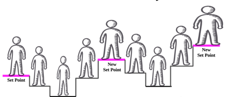
When you lose a significant amount of weight, the body does what it can to return to its original weight and the further from the original weight you drop, the greater the pressure to rebound.  The body seems to have a functional weight set point that it defends irrespective of the extent of its internal stores of energy (in the form of fat).   The set point can be raised by factors that affect the body’s homeostatic mechanism of weight maintenance.   While this set point can be raised, it does not seem to be amenable to being lowered.

Take the example of someone who has gained weight due to pregnancy.  The new higher weight is encoded as a new set point and if they then try to lose weight below this set point, the body counters with adaptive physiological processes that evolved to defend itself from starvation. These include the following mechanisms.

  1. Thyroid function changes.  Reverse T3 increases, T3 and T4 decrease, effectively rendering you hypothyroid
  2. Muscle efficiency increases (improved muscle function per calorie, less wasted calories)
  3. Neuro-humoral changes:  leptin, CCK, peptide YY all DECREASE, thus removing inhibitory actions on ghrelin, increasing appetite
  4. Decrease in resting energy expenditure due to weight loss and less available metabolically active tissue
  5. Adaptive thermogenesis decreases resulting in reduced resting metabolic rate
  6. Decreased sympathetic tone (and increased parasympathetic tone) result in less fat mobilization, slower physiology.

The result of these processes is that it is increasingly hard to maintain the new lower weight.  As a consequence, fewer than one out of six people who have lost a significant amount of weight can keep it off after a year.  The forces that return weight to the set point can be opposed by bariatric surgery and by anti-obesity medication.

There is a concern that by weight cycling, you end up resetting your basal metabolic rate lower across all weights.  So even after you regain the weight, you are still burning fewer calories, even at rest.  Why might this happen?

With weight regain, the body’s imperative can be understood as follows:  it seems to want to reconstitute fat free mass (FFM).  This includes lean muscle but also the weight of organs, bones and other non-fatty tissues in the body.   In Keys’ famous Minnesota weight loss experiment with conscientious objectors to WW2, over-eating and weight gain did not abate until FFM was replenished.  During weight regain, there was a desynchronization in the reconstitution of fat mass and fat free mass, so subjects ended up with an overshoot of fat return.  What this means is that when you regain the weight, you end up with an increased percentage of fat as compared to muscle.  Fat is a less metabolically active tissue and so you should theoretically have a lower BMR at the new weight, and this is what is observed.  So to be clear, not only do you regain weight, you end up with a slower metabolism for having lost it in the first place.

So consider the typical dieter who, while losing weight, will lose both fat and FFM (muscle, for our purposes).  As they regain the weight, they will regain primarily fat.  So functionally, they are replacing muscle with fat.  Not all their muscle, but enough that it affects their BMR.


Where does the set point reside?

How is this set point encoded and where does it reside in the body?   This turns out to be an enormously complex issue that is informed by the multiple variables that influence appetite including hormone signaling, homeostatic networks in the midbrain, bioenergetics related to mitochondrial metabolism, genetics, developmental epigenetic factors, the food reward circuits in the striatum, physical activity, and larger factors such as stress and socioeconomic forces.  This is a long way of saying we don’t know.  But let’s consider three theories.

  1. Leptin resistance:  the adipocyte-secreted hormone leptin increases in proportion to the amount of body-fat.  Reduction in body fat reduces the amount of circulating leptin, which triggers feeding behavior.  Conversely, an increase in leptin does not trigger reductions in intake unless the organism is leptin deficient.  Therefore leptin may represent a mechanism to protect against fat loss only.  Supplementing with leptin has not been shown to be a valid weight loss strategy.
  2. Maybe there is a ponderstat–an actual sensor in the hypothalamus.  Possibly more specifically, in the arcuate nucleus of the hypothalamus, where the major nuclei relevant to maintenance of weight reside.  One theory is that specific astrocytes in the arcuate nucleus either sense changes in nutrition or somehow are attuned to loss of weight, possibly in relation to leptin levels.  Alternatively, over nutrition induces an inflammatory reaction that changes neuronal function and resets the homeostatic system (as in the figure above).  Increased energy stores are encoded in a process known as reactive gliosis.[i]
  3. Mitochondrial theory: we know that mitochondria are irrevocably degraded by obesity.  Oxidative damage associated with obesity damages them, reducing their effectiveness and their numbers.  As a consequence, metabolism slows.  The consequence of widespread mitochondrial dysfunction, metabolism has been functionally reset because we are not using as much fuel, we cannot use it because we don’t have the mitochondrial capacity.  So with diminished energy use, there is a trend toward defending a higher weight.
  4. Changes in the microbiome:  we know that dietary restriction changes the microbiome and that those changes affect the degree to which the gut absorbs fat or lets it pass through the enteric tract.  Changes in the microbiome composition predispose (at least mice) to increased regain of fat after dieting.  There does not appear to be clear evidence yet in humans.

But then how do we keep the weight off?

  1. Bariatric surgery uniquely evades some of the post-weight loss changes related to a set point, but only for a time and then not completely.  The commonly performed surgeries change the composition and concentration of bile-acids, which are important signals of satiety.  Also, because of structural changes in the gut, more nutrition passes to the distal part of the intestine causing increases in anorexigenic hormones such as GLP1 and PYY (hindgut hypothesis).  There may also be changes in direct signaling to the CNS.  Then the mechanical issues: the stomach is smaller, there is a fear of dumping syndrome if you eat too fast, etc.  All of these factors contribute to a functional reduction in the set point, at least temporarily.  Yet many people who achieve weight loss after bariatric surgery subsequently regain some or much of the weight.
  2. Pacing. One basic principle is that if you must lose large amounts of weight, then do it slowly so that the body gradually adjusts to a lower set point.    Losing weight more deliberately and pausing between plateaus seems to help people evade the set point phenomenon and thus maintain reduced body weight,[ii] though to be clear, this finding is anecdotal, controversial and lacks experimental proof.
  3. Muscle.  When you lose weight, you lose both fat and fat free mass.  Fat free mass includes the weight of organs and other tissues, but also muscle.  When you regain the weight in the context of increased muscle mass, you will regain fewer pounds of fat.  In other words, muscle mass will protect against fat regain.  There are medications that should be used to mitigate the set point or even reset it. Ultimately, weight loss needs to be done in a controlled fashion with a plan, with frequent pauses to permit the body to catch up.  Muscle mass facilitates this process.
  4. Dietary characteristics. I suggest a weight loss diet that is somewhat less palatable, with less sugar, salt, fat and calorie density, more fiber.  The diet will be satiating but less rewarding.  If you can stick to a diet like this for a few weeks, it will change the brain reward centers and alter how you defend adiposity.
  5. Mitochondria: theoretically increasing the health and number of mitochondria will increase resting energy expenditure and may reset an elevated set point for the rationale suggested above. This would be done by zone 2 exercise and a variety of supplements that support mitochondrial biogenesis.  Also mitophagy inducers such as fasting and rapamycin.  In the future, maybe mitochondrial transplantation will be possible.
  6. Rapamycin: speaking of the namesake mtor inhibitor, it has been shown in rats to durably reset the ponderstat by unknown mechanism.
  7. Microbiome:  there is animal evidence that referring with a high protein concentration mitigates post weight loss fat regain.[iii]
  8. Finally, no discussion of weight is complete without a mention of exercise, sleep and stress management, the famous lifestyle triumvirate.  Changing these often seems so inaccessible to people who are locked in patterns of work, parenting and life-responsibilities.

[i] https://www.ncbi.nlm.nih.gov/pmc/articles/PMC6977167/pdf/main.pdf

[ii] https://www.nature.com/articles/ijo2017224

[iii] Zhong, W., Wang, H., Yang, Y. et al. High-protein diet prevents fat mass increase after dieting by counteracting Lactobacillus-enhanced lipid absorption. Nat Metab (2022).

Also https://www.sciencedirect.com/science/article/abs/pii/S001650851730152X