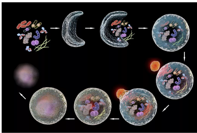
Autophagy

Autophagy (literally “self-eating”) is the body’s way of cleansing cells by recycling old or damaged components and is a process that appears to have a strong relationship to preventing disease and aging.  More technically, it is a highly regulated lysosome-dependent catabolic program used by the body to clear dysfunctional proteins, organelles and other structures that are typically first marked for the process by a protein called “ubiquitin.”  Primarily intra-cellular but also extra-cellular components are engulfed in autophagocytic vacuoles and degraded to constituent molecules, such as sugars, fatty acids and amino acids, which can be then be used as nutrition to produce ATP and/or provide building blocks for the synthesis of essential proteins.   There are different types of autophagy but for the most part they all serve this same purpose.  Mitophagy is a similar process that removes damaged or dysfunctional mitochondria.  There is also nucleophagy of material in the cell nucleus.


A number of types of autophagy are currently described.

  • Microautophagy: direct engulfment of cytoplasmic materials by invagination of lysosomal membrane.
  • Macroautophagy:  classic autophagasome mediated double membrane engulfment.
  • Chaperone Mediated autophagy: extremely selective target protein mediated (Hsc70) recycling that is activated in response to cellular stress.
  • Mitophagy:  autophagy related to mitochondria and their components
  • Also… ribophagy, lipophagy, xenophyagy, nucleophagy, clockophagy, lysophagy, etc.


Why is it important?
Autophagy is critical to cell function, regeneration and keeping the cell working properly.  Inhibition of autophagy by knocking out any of a variety of autophagy related genes invariably accelerates aging and disease.  Similarly, with aging, autophagy is downregulated (particularly in lymphocytes) leading to a forward feeding loop of accumulation of cellular debris and cellular dysfunction / dysregulation.   In contrast, enhanced autophagy delays aging and prolongs lifespan in a range of model organisms including yeast, nematodes, fruit flies and mice.  Lifespan studies in humans have not been conducted given the obvious challenges in conducting experiments in long-lived organisms, but it is generally assumed that regular induction of autophagy is a salubrious process, something we want to promote.  So how to do so?

How to promote autophagy?
First the biochemistry–skip ahead to the punchline if you prefer.  ULK1 is the enzyme that catalyzes production of phagosomes implicated in autophagy.  AMPK and SIRT activate ULK1, MTOR deactivates it.  If MTOR1 is inhibited by rapaymycin or starvation, autophagy is activated.  TFEB is considered the primary transcription factor for the synthesis of both lysosome and autophagosomes.   Again, AMPK boosts the activity of TFEB while MTOR inhibits it.  Spermidine, which declines with age, is a required co-factor in the translation of TFEB, so its supplementation is theorized (and shown) to stimulate autophagy.

Caloric restriction activates autophagy by stimulating AMPK and sirtuin1 as well as inhibiting mTOR.  When an autophagy related gene is knocked out or knocked down, the life-prolonging effect of rapamycin disappears, suggesting that autophagy plays an important role in the life-prolonging effect of rapamycin.

Simply not eating for a few hours and reducing nutrient levels in the blood starts the process of autophagy.  This is the rationale for time restricted feeding.  Starvation for longer periods such as two- or three-days initiates chaperone mediated autophagy, a targeted deep clean that modulates diverse functions such as glucose and lipid metabolism, DNA repair, cellular reprograming and the cellular response to stress.

Drugs to stimulate autophagy

  • Berberine and metformin both boost AMPK, thus phosphorylating and activating ULK1–> autophagy.
  • Rapamycin and spermidine inhibit MTOR, activating autophagy and spermidine, as already mentioned, both blocks motor and also is a necessary cofactor in the translation of TFEB.
  • Resveratrol and NR (nicotinamide riboside) theoretically target SIRT1, though evidence of their bioavailability after oral administration is questionable.
  • Ferulic acid may up-regulate SIRT1 expression and is derived from Angelica sinensis or “female ginseng” and used in traditional Chinese medicine.  It is also widely available as an additive in cosmetic skin creams.  Anthrocyanins in the diet are converted to ferric acid, providing a rationale for their health value.
  • Urolithin-A, a phytochemical derived from pomegranate has also been shown to boost SIRT1, as well as improve mitochondrial biogenesis.
  • Aerobic exercise has also been shown to stimulate autophagy.
  • It turns out these are all the usual suspects in the realm of anti-aging drugs and supplements, and while many of them have diverse actions, it may be that their action on the autophagy pathways is primary to their purported value as anti-aging interventions.  Yet the effect sizes of these interventions are not known, which is to say that we don’t really know to what extent autophagy will be triggered by taking any of these.

Can there be too much autophagy?  Perhaps yes.  While normal or even supernormal levels of autophagy promote cellular health, excessive or uncontrolled autophagy can initiate a type of cell death known as “autosis” (https://www.nature.com/articles/cdd2014143).  While autophagy is believed to be helpful, cell death through autosis is inarguably harmful and to be avoided.  The clinical significance of autosis in the context of therapeutic autophagy is not clear.  I reached out to Beth Levine, the woman who did much of the original research on autosis but her email bounced back and it turns out she has tragically died of breast cancer.  In any case, in this setting of incomplete understanding, the best course is one of prudence and moderation.  In the words of Hippocrates, first do no harm.  Practically speaking, my policy is to cycle agents, to avoid excessive dosing and to carefully follow serologic markers during treatment.

Relationship between autophagy and weight?
There is an emerging understanding of a relationship between autophagy in the medial-basal-hypothalamus (MBH) and obesity mediated by the FK506 (tacrolimus) binding protein FKBP51, which appears to be a major upstream regulator of autophagy.  Could autophagy and more broadly, the intracellular environment in hypothalamic neurons be the functional memory that induces phenomena such as the physiologic set point?  Could agents that augment autophagy such as rapamycin reset the physiologic set point in humans, as it has in mouse models?  These are intriguing questions.

Epigenetic clocks and Reprogramming  

How do we measure aging?  Certainly, there is the passage of time and how many birthdays someone has had, but we know that aging proceeds more quickly for some people than it does for others.  The speed of aging depends on your genes but also various factors such as, for instance, whether you smoke, your diet and level of exercise, your stress levels, exposure to ionizing radiation, and many other variables.  A 50 year old executive will likely be biologically younger than a 50 year old homeless alcoholic who has had a hard life, to take an example.  Is there some other marker that indicates a biological age that is distinct from chronological age?  This question is critically important if we are to assess the effectiveness of anti-aging interventions and has been a matter of much debate.  Recently as our understanding of aging has evolved, scientists have focused on methylation patterns in the DNA as a likely proxy for aging.

These so-called methylation clocks presuppose a process of epigenetic aging in which the organism’s DNA is gradually ornamented with methyl groups and that this methylation pattern is a good indicator of how old someone really is.  These methylations happen at points in the genome where cytosine is bound to guanine through a phosphate group (CpG).  When these CpG sites are methylated, it changes the three-dimensional structure of the DNA and changes the way that DNA can be expressed.  The theory holds that the loss of function with aging happens because the heavily methylated DNA is shaped differently and cannot be read (or transcribed) as easily as it once was.

These methylation patterns were discovered using deep computer learning analysis of tissue samples, by Steve Horvath and others made possible by affordable array-based technologies and massive amounts of publicly available human methylation data.  Horvath was able to identify 353 CpG methylation sites that predicted the progression of aging from embryonic stem cell to old age.   Of note, this is a very small fraction of the total number of CpG sites, which is itself a small fraction of the other methylations, histone acetylations and other changes that happen with age, so keep in mind that this is a highly focused view on a a process that is immensely more widespread.  Since his pioneering work, many different age clocks have been described which employ a variety of clusters of CpG methylation sites.  It’s not clear if these clocks are measuring the same thing[i].  In fact, the different clocks may be measuring different biological processes.  And this makes sense, since we know that aging proceeds at different rates across different physiologic systems, it’s not going to be a question of a particular clock as it is of multiple clocks, ticking independently.  How do you assess aging in different tissues like the brain?  At this point, genetic material is not sampled from each tissue—nobody is doing brain biopsies— but rather only from white blood cells, and then methylation sites are assigned to tissues based on the type of protein coded by the DNA region.

Are these methylation patterns a marker for aging or are they actually driving aging?  This is not a settled question.  We do know that increased epigenetic age relative to chronological age has been linked to disease and mortality risk.  And removing methylations in mouse retinal cells (using Yamanake factors, see below) and reintroducing these cells into the retina using a viral vector reversed vision loss and cured blindness in mice.[ii]   The researchers believed that they were reprogramming the cells to an earlier (younger) state of their progression on the program of methylation.   Reprogramming means taking aged or otherwise faulty cells and tissue that have already grown and “differentiated,” and resetting them closer to an original state of induced pluripotency.  This is done using a group of transcription factors called Yamanake factors, discovered by the Nobel laureate Shinya Yamanaka.  These Yamanake transcription factors regulate the expression of DNA in such a way that the cell returns to a state where it can become any kind of tissue.  They are:

Oct-3/4:  transcription factors critical for maintaining pluripotency and preventing cell differentiation
Sox family:  important for sex determination but also maintenance of pluripotency
KLF4: regulation of apopotosis, tumor suppression
c-Myc: proto-oncogene

One of the actions of the Yamanke factors is to make possible the de-methylation of the same CpG groups that are theorized to drive differentiation and aging.  Myc is sometimes left out because it is associated with uncontrolled growth, leaving Oct/Sox/KLF aka “OSK” treatment.  OSK treatment in mice has been shown to reduce levels of methylation and also to restore function.  If reprogramming with OSK is pursued to its natural conclusion, cells return to pluripotency and can become cancerous.  However, if the reprogramming is done incompletely, then the cell simply becomes younger.  This is the aim of cellular reprogramming in a nutshell, and an enormous amount of money is being invested in this field ($3 billion for Bezos funded Altos labs last week) to capitalize on the technology.  The goal will be to safely do partial reprogramming, to avoid pluripotency and the risk for malignancy.  In a recent paper, they appear to have done just that with mice.

What is driving the methylations?  Is it just DNA damage, a stochastic and random process, as David Sinclair has proposed?  Or is it some programmatic process from birth to death?   We don’t know yet.  One observation supporting randomness is that methylation changes are more likely to occur at early and late replication sites in DNA.  In other words, when a cell divides and DNA is copied to create two daughter cells, the epigenetic changes are also copied.  The observation that the epigenetic changes are concentrated at specific sites relevant to the replication process suggests some mechanical issue rather than an external program driving methylation from embryogenesis to old age.  On the other hand, how is would it be that all cells in the body (or at least a statistical majority) should change their epigenomic methylation patterns simultaneously?  The improbability of this occurrence argues for a central regulator.  Thus far, empiric evidence either way is scant.

Another question is whether reprogramming creates a truly rejuvenated cell in every sense of the word.  We know treatment with Yamanake does not remove all methylations, but only important subsets of them. Is there longevity for these cells?  How do they behave over time.  These are questions that remain to be resolved.  Hopefully in the next decade we will see clinical applications that derive from this technology in human beings.  It will be expensive at first, but I’m sure at least one child with a previously incurable disease will be granted a normal life thanks to reprogramming.  And he or she will be free to age normally and watch the world gradually warm to uninhabitable levels and descend into geopolitical chaos, but that is a matter for another blog post.

[i] https://onlinelibrary.wiley.com/doi/epdf/10.1111/acel.13229

[ii] https://www.ncbi.nlm.nih.gov/labs/pmc/articles/PMC7752134/pdf/nihms-1640389.pdf

One way of looking at anti-aging interventions

The hallmarks of aging as elaborated in the now famous paper are the key biological changes that occur in living organisms as they grow older. These changes are thought to be the underlying cause of many of the health problems associated with aging, such as loss of muscle mass, declining cognitive function, and increased susceptibility to disease. The nine hallmarks of aging are:

  1. Genetic instability – accumulation of mutations in the DNA
  2. Telomere attrition – shortening of the protective caps on the ends of chromosomes
  3. Epigenetic alterations – changes in gene expression that do not involve changes to the underlying DNA sequence
  4. Loss of proteostasis – decline in the cell’s ability to maintain the proper folding and function of proteins
  5. Deregulated nutrient sensing – changes in the way cells respond to nutrients and growth signals
  6. Mitochondrial dysfunction – decline in the function of the mitochondria, the cell’s powerhouses
  7. Cellular senescence – accumulation of cells that can no longer divide and perform their functions
  8. Stem cell exhaustion – loss of stem cells and their regenerative abilities
  9. Altered intercellular communication – changes in the way cells communicate with each other.

These changes are thought to contribute to the aging process and the development of age-related diseases.

Of course, these hallmarks have raised the very important question of whether there is one process to rule them all.  Do all of these hallmarks accumulate with the passage of time or are they programmed in some way.  That is the question and at this time there are only theories.

Attempts at “treating” or forestalling aging, focus on these hallmarks.


1. MTOR inhibition.

MTOR (or mechanistic target of rapamycin) is a highly conserved kinase (or enzyme) present in all cells in almost all forms of life. It’s purpose is to sense whether or not the organism is in a fed state, and if so, it signals cell growth and expansion in numerous ways. Conversely, if the enzyme senses a lack of nutrition, it puts the cell in a state of frozen animation that theoretically permits it to persist until the time that nutrition is readily available. Supporting this theory of aging, calorie restriction with adequate nutrition has been shown to extend lifespan in a wide range of life forms: yeast, worms, flies, mice and monkeys. But not yet humans, because it’s very hard to do lifetime experiments to test this in people. Even so, there is plenty of evidence that reducing calories decreases metabolism, reduces oxidative damage to tissues and profoundly changes the body in lasting ways. The challenge is to derive these benefits without resorting to severe caloric restriction, which is widely considered not fun.

2. Cellular senescence

Cells can enter a zombie state called senescence in which they no longer grow or divide but they take up space and even secrete inflammatory factors into the local environment that persuade other cells to become senescent. They can be detected histologically by stains that turn the senescent cells blue, which allows us to see the profound burden of senescence in older individuals. Senescence may have some utility to the organism in facilitating scarring and perhaps other necessary roles, but removing senescent cells has been shown to have remarkable rejuvenating effects in older lab animals. A number of companies are banking on the theory that we will oneday treat disease or rejuvenate older individuals with senolytic medications.

3. Mitochondrial aging

Mitochondria are the engines of the cell that provide us with energy. They are likely foreign bacteria that were captured into a symbiotic relationship early in evolution, that have been incorporated into our cells and are not virtually indistinguishable from their hosts (other than having separate DNA). As we age, they age as well and we perceive this decline in number and function as a diminishment of energy. Are there ways to rejuvenate mitochondria? The answer is probably. Certainly exercise, and in particular zone 2 exercise, has been shown to improve mitochondrial function. Beyond that there are supplements that have been shown to support mitochondrial function, especially in old age, with resulting improvements in cognition and muscle strength.


4. Epigenetic changes / Reprogramming

We are born with certain genes that do not change, but which genes are used or expressed changes over time and in response to the conditions of life.  Epigenetics refers to changes of gene expression that happen without changes to the DNA coding sequence.  These changes are put into action by changes in DNA methylation and acetylation patterns, modification of histone, and chromatin remodeling.

Methylation (or acetylation) of DNA by repair enzymes changes the proper analog folding and orientation of DNA chromatin strands in the nucleus leading to epigenetic phenotypic changes. We age because our cells forget the original intentionality that is encoded in the epigenome. Basically, youth → living → broken DNA → genome instability → disruption of DNA packing and gene regulation (the epigenome) → loss of cell identity → cellular senescence → disease → death.  Steven Horvath and others have analyzed these methylation patterns and their predictable progression and claim to be able to use them as a biomarker for aging. You can pay to get your biological age typed with a Horvath clock. Or a Grim clock or other clocks. The undoing of this methylation could theoretically reprogram cells and rejuvenate organisms and there is some experimental evidence that it works, but this is seemingly far from widespread application or availability.  Some lower life forms (sponge, hydra) have access to the germline that can reset methylation to an earlier state.  According to this theory, they should be immortal.  And it turns out that they are.  Sinclair and others believe that germline information can be reintroduced into somatic cells by way of adenovirus vectors to restore youthful phenotype.

**In less than a year, this technology seems closer to fruition.